
A step forward in understanding hydrocephalus
Hydrocephalus is a devastating structural neurological disorder marked by enlarged brain ventricles due to accumulation of cerebrospinal fluid.
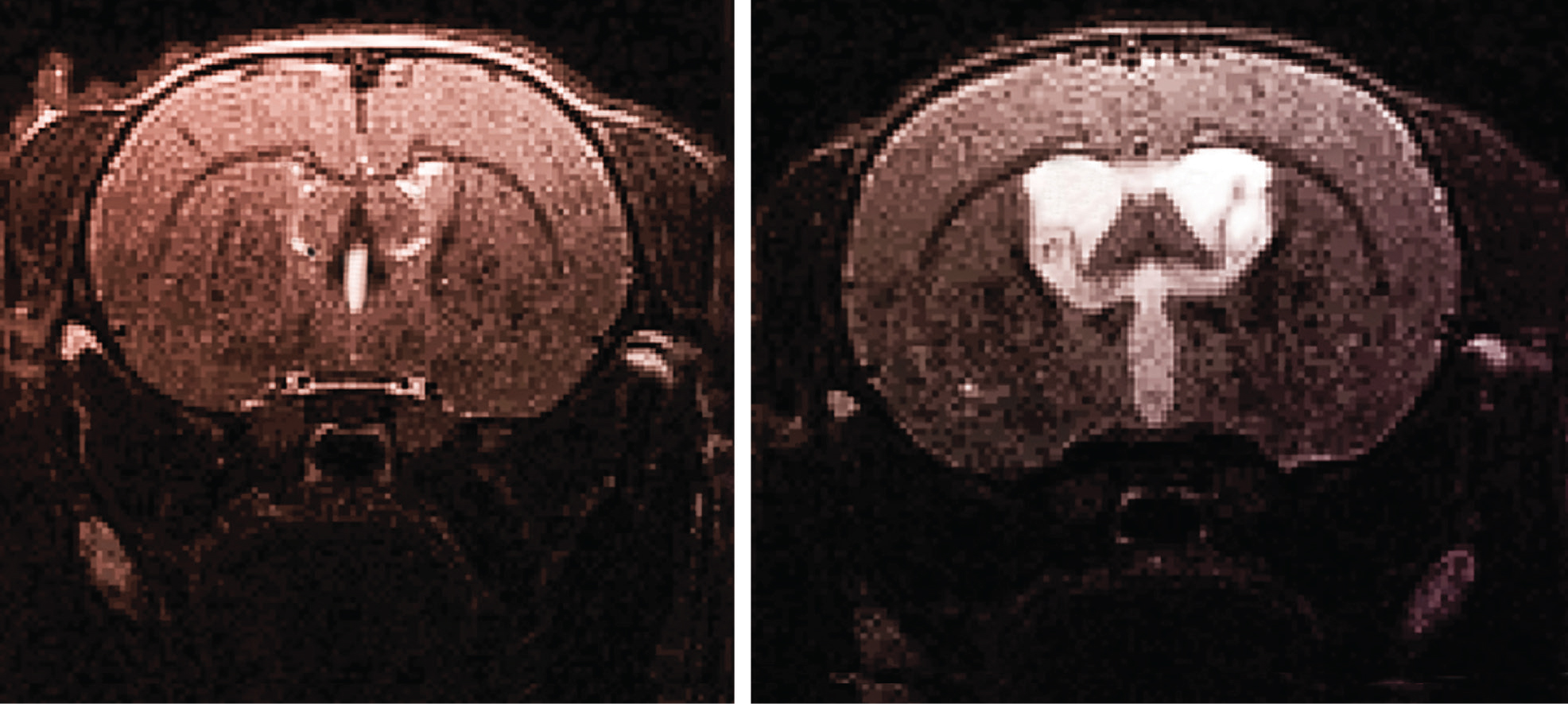
High resolution anatomical images of a normal (on the left) and hydrocephalic (on the right) rat brain acquired using Magnetic Resonance Imaging
The current diagnosis and treatment of hydrocephalus is inadequate due to a lack of understanding about the mechanisms behind its development.
Hydrocephalus may be accompanied by low intracranial pressure and it continues to remain a clinical challenge to differentiate this disease with other diseases such as cerebral atrophy based on ordinary radiography images.
While hydrocephalus can be treated with shunt, similar treatment on cerebral atrophy could be fatal. By using novel imaging technology, our team is tracking how important physiological factors in the brain change during hydrocephalus. Our goal is to identify new imaging biomarkers that can help us with diagnosis and prognosis. For example, change in brain tissue stiffness has been widely known to play an important role during the development of the condition.
Over the past few decades, scientists have studied this by measuring brain tissue stiffness in vivo, which requires a craniotomy. This procedure is invasive and brain tissue stiffness measured using this method is limited to the superficial layer and is likely to include stiffness of the dura and other brain tissue structures.
In a world first, by using Magnetic Resonance Elastography, our research team has characterised how brain tissue stiffness changes during the development of hydrocephalus non-invasively and in the deeper brain regions.
This unique finding paves the way to develop stiffness indices that can be used to guide treatment prognosis thereby improving the clinical management of patients.